
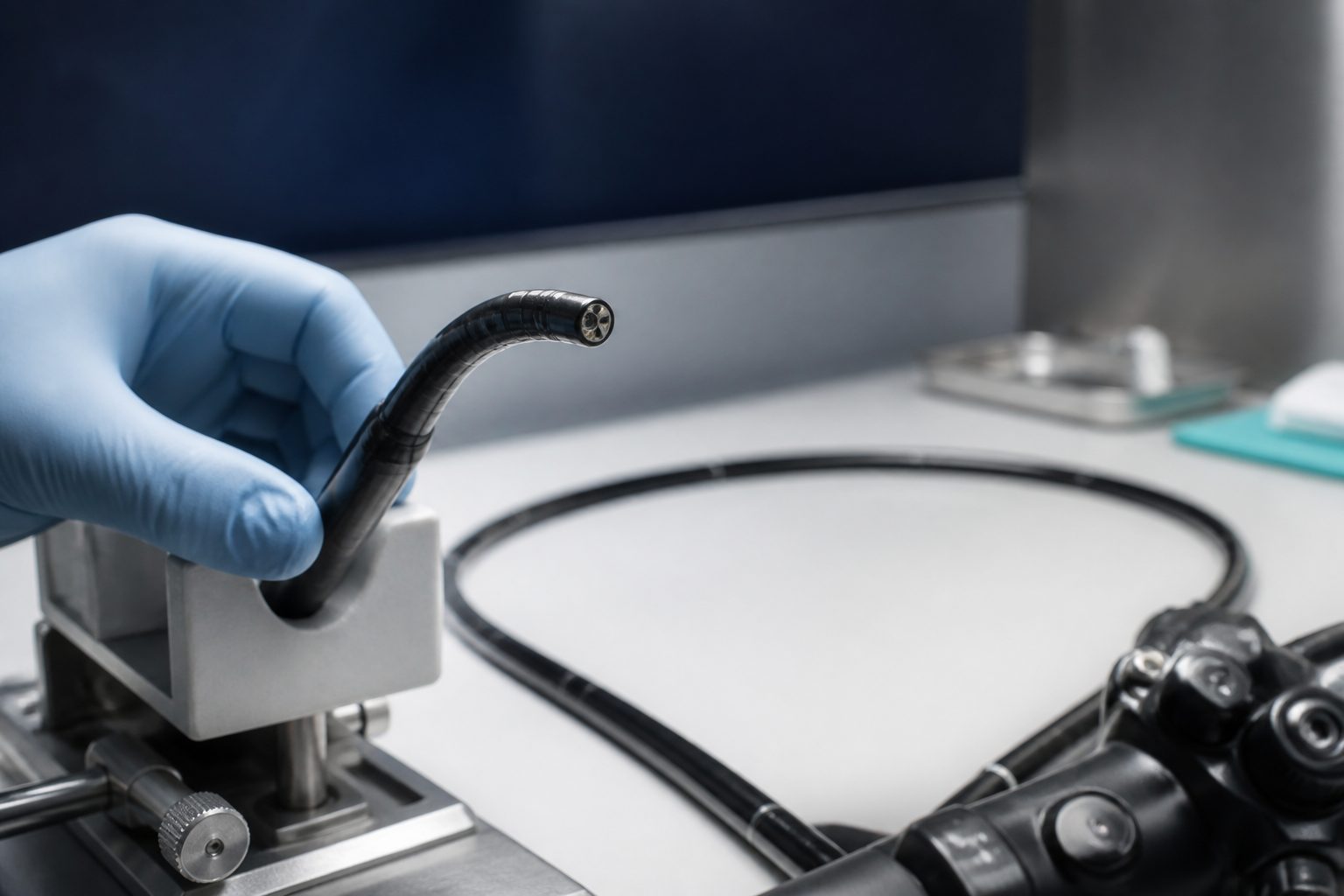
ENT
Rhinolaryngoscope Repair for ENT Clinics and Practices
ENT scopes take their damage between cases as often as during them. We repair image bundles, distal tips, angulation, and sheaths on both fiberoptic and chip on tip rhinolaryngoscopes, with a written quote before any work.
- Image bundle and broken fiber repair
- Distal tip and objective lens work
- Angulation and sheath restoration
- Fiberoptic and chip on tip scopes
Get a Free Repair Quote
Tell us what failed and we will send a written quote before any work begins. No charge for the evaluation, and no purchase order needed to get one.
Quote before any work
You approve a written quote before a technician touches the scope.
Free evaluation
We diagnose the failure and price the repair at no charge, even if you decline.
We come and get it
Pickup and delivery across Southern California, mail-in service everywhere else.
Independent, not the OEM
All major manufacturers serviced, without OEM exchange pricing.
By symptom
Rhinolaryngoscope Failures Seen in ENT Clinics
Most of these build up quietly in a clinic drawer. Find the one on your bench and call with the model number.
Broken fibers in the image bundle
Each fractured fiber prints a permanent black dot in the field. They accumulate from bending, dropping, and pressure on the shaft, and the count only rises. Bundle repair or replacement is what brings the field back.
Broken illumination fibers
The light carrying bundle fails the same way the image bundle does, a few fibers at a time. Turning the source up masks it for a while. We test the illumination path separately from the imaging path.
Distal tip and objective damage
The tip contacts teeth, nasal bone, and drawer edges, and a chipped objective window scatters light across the whole field. Tip and lens work restores contrast rather than just brightness.
Slack angulation wires
The lever moves further than the tip does, or the tip drifts back before the view is captured. Wires stretch on a thin ENT scope quickly once they start. Replacement and tension reset bring the travel back to spec.
Sheath damage on the insertion shaft
A cracked outer layer lets disinfectant into the shaft, which is how a cosmetic problem becomes an internal one. On a scope that is wiped and soaked between patients that transition is fast.
Chip on tip camera or cable fault
On a video ENT scope the imager, the internal cable run, and the connector each fail differently. We isolate the fault along that path rather than replacing the most expensive part first.
Black Dots in the Field and Image Bundle Fiber Breaks
On a fiberoptic ENT scope, the picture is carried by thousands of individual glass fibers packed into a bundle a couple of millimeters across. Every fiber is one pixel. When a fiber fractures it goes dark permanently, and it never comes back, so the field slowly fills with black dots that no cleaning and no light source adjustment will remove.
The breaks come from bending the shaft tighter than it was built for, from dropping the scope, and from anything that puts point pressure on the insertion section, including a drawer that gets closed on it. Clinics tend to tolerate the dots until a specific number of them lands in the middle of the view. By then the bundle usually needs replacement rather than repair.
The useful window is earlier. A count that is climbing is telling you something about handling and storage, not just about the scope, and a device evaluated at that point is a much smaller job. Every scope we take in goes through the same intake as the rest of our flexible endoscope repair work.
Chip on Tip Scopes and Distal Tip Damage
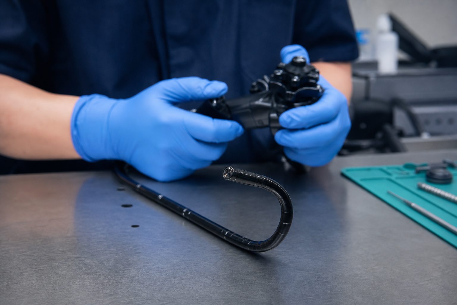
Video ENT scopes moved the imager to the distal end, which removed the fiber bundle and replaced it with a small camera, an internal cable run, and a connector. That trade takes away the black dot problem and introduces three new failure points, each of which produces a dropout or a flicker that looks the same on the monitor.
The distal end still takes the same physical abuse either way. Contact with teeth during a laryngoscopy, a knock against nasal bone, and drawer edges all chip the objective window, and a chipped window scatters light across the whole picture rather than blocking one part of it. That specific pattern is covered on our distal lens and objective page.
We isolate faults along the video path instead of swapping the costliest component and hoping. A connector fault and an imager fault produce similar symptoms and very different quotes.
How ENT Scope Storage and Handling Shortens Service Life
These devices live differently from hospital scopes. They sit in exam room drawers, they are wiped and soaked between patients rather than run through a central reprocessor, and they are frequently handled by staff who were never trained on scope care because the clinic never had a sterile processing department to do that training.
The result is a predictable damage pattern. Cracked outer sheaths from disinfectant exposure through an existing scratch, bundle breaks from tight coiling in a drawer, chipped tips from contact with other instruments, and slack angulation from a lever that has been worked hard. None of it happens during a procedure, which is why it is rarely reported as an incident.
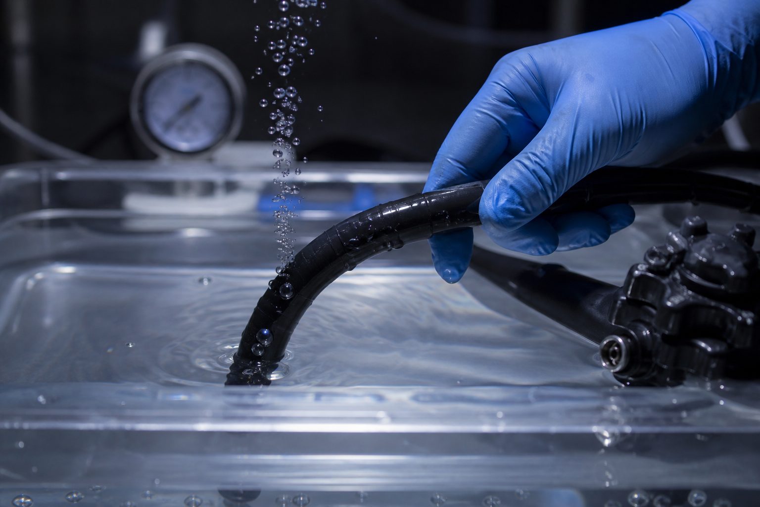
A cracked sheath deserves attention first, because that is the fault that lets fluid into the shaft. If a device has already taken fluid internally, our fluid invasion page explains what is recoverable, and a scope that will not hold pressure belongs on the leak testing page before anything else.
Rhinolaryngoscope Optics, Angulation, and Sheath Faults Together
Most ENT scopes that reach us arrive with more than one problem, because nothing prompted a look until the image finally became unusable. A dim field, a chipped tip, and a lever that no longer holds deflection are three separate repairs that share a single cause, which is a device nobody inspected for two years.
You receive the full condition report and decide what to authorize. Practices frequently approve the optics and defer the angulation, or the reverse, and both are sound choices once the whole list is in front of you. If deflection is the priority, the work is described on our steering and angulation page.
Most of these scopes carry a Pentax Medical ENT nameplate or an Olympus label, and we are independent of both. Clinics that also run other flexible devices send them together, so a pickup may include a bronchoscope with a blocked channel or a gastroscope with a scratched lens.
How it works
How Rhinolaryngoscope Service Works From Pickup to Return
A free evaluation, a written quote you can act on, and no work until your practice approves it.
Tell us what failed
Send the manufacturer, model, and what you are seeing. A photo of the damage helps but is not required.
We collect the scope
Courier pickup across Southern California, or ship the device in to our bench from anywhere in the country.
You get a written quote
We evaluate the device, document the failure, and price the repair. Nothing proceeds until you approve it.
Repaired, tested, returned
The device is repaired, leak and function tested, and returned with documentation for your records.
Questions we get
Rhinolaryngoscope Repair Questions From ENT Practices
Can the black dots in our image be removed?
Our clinic has no sterile processing department. Does that change anything?
Do you work on video scopes as well as fiberoptic ones?
The outer sheath is cracked but the picture is fine. Is that urgent?
How do we get the scope to you from a clinic?
Other Flexible Scope Repair We Provide
The rest of the flexible fleet goes through the same bench and the same intake.
Get a Free Evaluation and Rhinolaryngoscope Service Quote
Send the manufacturer, the model, and what the image looks like. We price the repair before anyone opens the scope.
Monday to Friday, 8:00 AM to 5:00 PM Pacific
Saturday and Sunday, closed
Mail-in repairs received any day
Serving Southern California with pickup and delivery, and the rest of the country by mail-in.